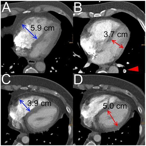
rv lv ratio The RV/LV ratio is determined by measuring the maximal RV and LV diameters from inner wall to inner wall on the axial slice that best approximates the four-chamber view (Fig. 9) . A value > . Ideal for locations requiring cable bending. Flexible stranded center conductor. High-density braided shield. Dielectric strips away clean from conductor. LV-61S is equivalent to RG-59B/U. Stays flexible in sub-zero weather Note: Cable strippers (TS100 series) cannot be used for L-1.5C2VS and LV-77S
0 · rv vs lv failure
1 · rv lv ratio pulmonary embolism
2 · rv lv ratio on ct
3 · rv lv ratio measurement
4 · rv lv ratio meaning
5 · rv lv ratio calculator
6 · right ventricular spiral of death
7 · normal rv to lv ratio
Now though, everything has changed. It's like the Devs just decided to forgo linear progression after level 30 altogether and just dumped all the missions at once. I'm a PVE player that's invested in story content, but now it's all jumbled and it's hard to tell where missions go chronilogically.
According to the latest European Society of Cardiology (ESC) guideline, a right ventricle–to–left ventricle (LV) diameter ratio >1.0 is the most appropriate method for determining dysfunction .
The right ventricular to left ventricular diameter (RV:LV) ratio measured at CT pulmonary angiogram (CTPA) has been shown to provide valuable information in patients with pulmonary arterial hypertension and to . This technology has been tested in randomized controlled trials using the endpoint of improvement in RV/left ventricular (LV) ratio because this predicts mortality and adverse outcomes. 17 Safety endpoints include major .The RV/LV ratio is determined by measuring the maximal RV and LV diameters from inner wall to inner wall on the axial slice that best approximates the four-chamber view (Fig. 9) . A value > .RV/left ventricular (LV) ratio on CTPA is a clinical endpoint in clinical trials involving catheter-directed treatments in patients with intermediate-risk pulmonary embolism (7 – 11). Identifying .
Evaluation of the right ventricle (RV) is a key component of the clinical assessment of many cardiovascular and pulmonary disorders. There are many ways to . the right ventricular outflow tract is considered enlarged when the measured diameter in the parasternal long axis exceeds 3.3 cm, or when the measured diameter .

rv vs lv failure
RV dilatation as a proxy for RV dysfunction can be assessed by calculating the right-to-left ventricle diameter (RV/LV) ratio on standard computed tomography pulmonary . Functional studies of the right ventricle (RV) have been overshadowed by studies of left ventricular function for a long time; some investigators even concluded during the first half of the 20th century that . A ratio of systolic to diastolic duration <1.00 was associated . and septal displacement also create RV dyssynchronous motion 81 – 83 and dyssynchronous RV-LV contraction. 65,83,84 Delayed RV lateral wall .
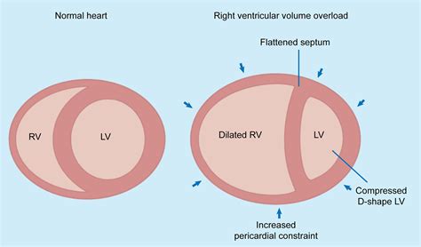
In contrast to the RV/LV diameter ratio <1 group, RV/LV diameter ratio >1 group showed significantly more number of scores 2 and 3 patients. Also, there were not any score 4 and 5 patients in RV/LV diameter ratio <1 group, .Patients with an RV/LV ratio of ≥1 had statistically significantly higher troponin levels (p= 0.004) and IVC reflux (p= 0.025) compared to patients with an RV/LV ratio of <1. Conclusions: In conclusion, RV/LV ratio should be evaluated together with cardiac biomarkers to . Echocardiography is the best imaging study to detect RV dysfunction in the setting of acute PE. Characteristic echocardiographic findings in patients with submassive PE include RV hypokinesis and dilatation, interventricular septal flattening and paradoxical motion toward the LV, abnormal transmitral Doppler flow profile, tricuspid regurgitation, pulmonary hypertension as . The RV/LV ratio was shown to correlate with invasive measurements of PH (12, 26) and incorporates leftward septal shift and RV dilatation, thereby incorporating RV failure, remodeling, and adverse hemodynamics. An RV/LV ratio > 1 has been associated with increased mortality risk and thus is clinically useful .
Higher RV/LV ratios increase specificity for decompensation (16–18) regardless of the patient’s hemodynamic stability. Therefore, RV/LV ratios of >1.0 should be used to risk stratify patients . This is as recommended in current clinical practice guidelines, as outlined in the 2019 European Society of Cardiology recommendations .
Where multiple RV/LV ratios were reported, we utilized the one with the highest sensitivity. The pooled sensitivity of increased RV/LV ratio was 0.83 (95% CI=0.78 - 0.87; I 2 = 82.9) while the pooled specificity was 0.75 (95% CI=0.66- 0.82; I 2 = 94.6) (Figure 1). Considering all RV/LV ratio studies, the summary receiver operating .RV/LV ratio calculator. Articles Cases Collections Templates Tools Search Assistant Dashboard Login RV/LV Calculator. RV Diameter: LV Diameter: The RV:LV ratio had poor specificity in detecting PH at RHC however, suggesting that the RV:LV ratio at CT cannot be relied upon to exclude PH. In our cohort, a high proportion of patients had PH (78%). Of 80 patients with an RV:LVlargest ≥1.0, 16 (20%) had borderline PH (mPAP of 21[16-23] mmHg, and PVR 2.8[1.8-3.1] Wood units). This may . In our previous work 5, in a group 152 controls, we determined the normal range of the RV/LV volume ratio to be 0.906 to 1.266, and found that combining this parameter (RV/LV volume ratio ≥1.27 .
RV/LV Analysis provides annotated images showing the ventricular measurements and a summary report of the RV/LV ratio, automatically delivered to the patient study in PACS. FDA 510(k) Cleared, CE Mark Pending. The Power of Personalized Quantitative Imaging. Outcomes of interest included the proportion of patients treated at home with a RV/LV ratio >1.0 and comparing the 3-month incidence of recurrent venous thromboembolism and mortality in patients with and without RV dilatation. Of the 1627 patients eligible for the study, RV/LV ratios were available for 1474 patients, of whom 752 were treated at .
In our study, RV/LV volume was more accurate than both uni-dimensional RV/LV diameter ratios (RV/LV axial and RV/LV 4-CH) for predicting echocardiographically confirmed RVD in patients with acute pulmonary embolism. The investigators concluded that an increased RV:LV ratio calculated at CTPA provides a simple, noninvasive, useful screening tool for risk stratification in patients with suspected ILD-PH. These findings should encourage closer follow-up, more aggressive treatment, and consideration of lung transplantation in these individuals. . The accuracy and reproducibility of RV/LV diameter ratio measurements by non-radiologist clinicians without dedicated training and expertise in CT reading is unknown. We aimed to assess the accuracy and reproducibility of CTPA RV/LV diameter ratio measurement by three residents in internal medicine without prior dedicated training in CT reading. TAPSE / PASP ratio may be a good marker of RV-PA coupling (or uncoupling) as TAPSE reflects RV contractile function and PASP is a surrogate for afterload TAPSE/PASP ratio normally >0.31mm/mmHg; A ratio of .
RV/LV ratios were determined in consensus by 2 radiologists with extensive experience in cardiovascular CT using standard axial plane reconstructions. Both readers were blinded towards sleep study results. Maximum RV and LV diameters were defined manually as the maximum distance of the endocardial border of the right or left ventricle from the .
Purpose: The purpose of our study was to determine if the ratio of right-to-left ventricular diameter (RV/LV ratio) on computed tomography (CT) pulmonary angiograms (CTPA) is predictive of 90-day mortality in patients without pulmonary embolism (PE). Materials and methods: This Institutional Review Board-approved single-institution retrospective study was .So normally the RV:LV ratio should be about 0.6 to 1 and this is classically measured in the apical four chamber view across the tips of the valves. We choose 1:1 as being abnormal because if you're getting the view correctly, it’s definitely abnormal when the ratio exceeds 1, when the RV is equal to or greater than the LV.
The RV/LV ratio increases with GA, although without clinical significance. These reference values will be useful in objective assessment of RV-to-LV disproportion. Reference values for cardiac ventricle widths and their ratio throughout gestation were established. The RV/LV ratio increases with GA, although without clinical significance. An RV-to-LV ratio greater than 1 has a good correlation with echocardiographic detection of RV dysfunction [35, 36]. To get results more like those of echocardiography, it is possible to measure this ratio on a reformatted four-chamber view; a ratio greater than 0.9 shows a certain degree of correlation with morbidity and mortality .
Right ventricular enlargement (also known as right ventricular dilatation (RVD)) can be the result of a number of conditions, including:. pulmonary valve stenosis; pulmonary arterial hypertension; atrial septal defect (ASD) ventricular septal defect (VSD) tricuspid regurgitation
The RV/LV end-systolic diameter ratio can easily be obtained noninvasively in the clinical setting and can be used in the management of patients with PH. The RV/LV ratio incorporates both pathologic septal shift and RV dilation in children with PH and correlates with invasive measures of PH. An RV/L .
right ventricle/ left ventricle end diastolic basal diameter ratio >1. the right ventricular outflow tract is considered enlarged when the measured diameter in the parasternal long axis exceeds 3.3 cm, or when the measured diameter exceeds 2.7 cm in the distal RVOT, as measured in the basal parasternal short axis viewThe primary aim of this study was to assess the incidence of CT-measured RV dilatation, defined as a CT-assessed RV/LV diameter ratio greater than 1.0 with the ventricular diameters measured in the transverse plane at the widest points between the inner surface of the free wall and the surface of the interventricular septum, and its impact on clinical outcome. Aim: Increased ratio between the right and left ventricular (RV/LV) diameters ≥1 is considered an important imaging marker for risk stratification among patients diagnosed with acute pulmonary embolism (PE). Our goal was to assess the prevalence of RV/LV≥1 among consecutive patients undergoing computed tomography pulmonary angiography, and to .RV:LV ratios ranged from 0.67 to 2.43, with 64% (n = 65) > 1.0. There was very strong agreement between manual and automated RV:LV ratios (ICC = 0.83, 0.77-0.88). The use of automated analysis led to a change in risk stratification in 45% of patients (n = 40). The AUC of the automated measurement for the prediction of all-cause 30-day mortality .

rv lv ratio pulmonary embolism
Find out what you can do in your account, how to know if you already have one and what to do if you've forgotten your details. Find out more in our handy guide.
rv lv ratio|rv lv ratio calculator


